Trauma Management
When a patient receives trauma to their teeth, the management is very important in improving the success of treatment.
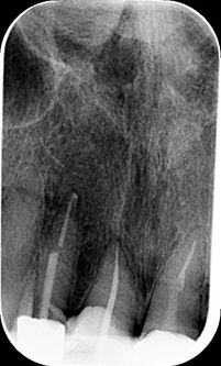
This young gentleman had an accident at rugby that displaced the tooth. The tooth was reinserted, a “flexible” splint applied and as the tooth died, a root canal filling using Mineral Trioxide Aggregate (MTA) was used.
Root Canal Retreatment
Root canal retreatment is needed when the original treatment has not worked and the tooth is usually giving symptoms. During the retreatment the old root canal material is removed. Quite often a reason for failure is identified and this can be a missed canal or that the original treatment is short of the optimum length.
Retreatment missed canal radix enteromolaris
Retreatment 5 canal molar
Missed canal
Lateral Canals
Lateral canals are small root canal offshoots from the main root canal. We use ultrasonic cleaning and disinfecting techniques to reach deep into these irregularities.
Case 1
Case 2
Case 3
Complex Primary Root Canal Treatment
For a root canal treatment to have the best possible chance of success, we need to find and clean all of the canals inside a tooth.
Complex molar anatomy
- extreme curvature
Some teeth are move curved than others. When a tooth has twisted roots it is more difficult to open and disinfect the tooth. We use a special protocol to open these along with the finest quality Swiss made instruments.
Complex molar anatomy
- case 2
Lower molars can have three, four or even five canals. The dental operating microscope allows us to search for these canals and clean and disinfect the tooth.
Complex molar anatomy
- case 3
Second mesiobuccal canal
A second mesiobuccal canal is present in 96% of upper first molars and 70% of upper second molars.
These are usually very small, however, the use of the dental operating microscope and specialised ultrasonic tips allows us to identify these and clean them.
Root Canal Obliteration
Root canal obliteration can occur after a tooth has received some trauma in the past. The tooth canal gets smaller over time, to the point it is microscoptic. Normally the tooth stay symptom free however if the tooth finally dies and needs root canal treatment it can be challenging to get into the canal system.
Post Removal
Case 1
Case 2
Perforation Repair
When a tooth is opened of root canal treatment the canals can be very small and difficult to locate. This can result in a dentist perforating through the side or floor of a tooth. These areas can be carefully repaired with mineral trioxide aggregate (MTA) and the tooth root canal treated as normal.
External Cervical Invasive Resorption
External invasive cervical root resorption is a condition whereby the cells that normally control bone turn over mistake the tooth dentine. The cells then destroy the dentine and replace the tooth with soft tissue. If left untreated the tooth is eventually lost.
CBCT
Teeth can have very complex internal anatomy. We can now use 3D x-rays called cone beam computed tomography (CBCT) to look at the root from different angles. This helps us diagnose more tooth problems and also helps guide our treatment.
Case 1
Case 2
Bioceramic Apexification
When a tooth has a large opening into the bone, the standard “gutta percha” rubber is no longer the ideal filling material. In these cases we use mineral trioxide aggregate (MTA) to seal the tooth. This is known as a “bioceramic” and the bone can grow onto its surface.
Case 1
Case 2
Apical Surgery
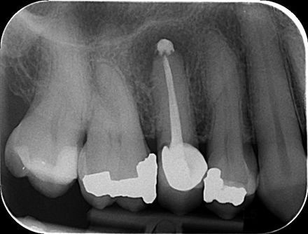
A tooth can be root canal treated from either the top (orthograde) or through the tip of the tooth (retrograde). We normally prefer to treat through the top of tooth however this does not allows work or sometimes there is an obstruction such as post and we decide to undertake a surgical procedure.
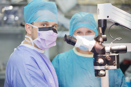
We perform microsurgery under the dental operating microscope using specialist instruments and materials. Robert has undertaken advanced training with one of the UK’s leading experts in area Dr Bhandari.
Broken Instrument Removal
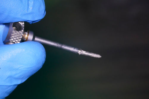
Robert has trained with one of the worlds best at removing fractured dental instrument’s “Dr Terauchi”
We use the dental operating microscope and special “micro” instruments to carefully remove these.


